Insight into ear growths
An abnormal skin growth in the middle ear behind the eardrum is called cholesteatoma. Repeated infections and/or and a tear or retraction of the eardrum can cause the skin to toughen and form an expanding sac. Cholesteatomas often devolop as cysts or pouches that shed layers of old skin, which build up inside the middle ear. Over time, the cholesteatoma can increase in size and destroy the surrounding delicate bones of the middle ear. Hearing loss, dizziness, and facial muscle paralysis are rare, but can result from continued cholesteatoma growth.
A cholesteatoma usually occurs because of poor eustachian tube function as well as infection in the middle ear. The eustachian tube conveys air from the back of the nose into the middle ear to equalize ear pressure (“clear the ears”). When the eustachian tubes work poorly, perhaps due to allergy, a cold, or sinusitis, the air in the middle ear is absorbed by the body, creating a partial vacuum in the ear. The vacuum pressure sucks in a pouch or sac by stretching the eardrum, especially areas weakened by previous infections. This can develop into a sac and become a cholesteatoma. A rare congenital form of cholesteatoma (one present at birth) can occur in the middle ear and elsewhere, such as in the nearby skull bones. However, the type of cholesteatoma associated with ear infections is most common.
An examination by an Otolaryngologist—Head and Neck surgeon can confirm the presence of a cholesteatoma. Initial treatment may consist of a careful cleaning of the ear, antibiotics, and ear drops. Therapy aims to stop drainage in the ear by controlling the infection. The growth characteristics of a cholesteatoma must also be evaluated.
A large or complicated cholesteatoma usually requires surgical treatment to protect the patient from serious complications. Hearing and balance tests, x-rays of the mastoid (the skull bone next to the ear), and CAT scans (3-D x-rays) of the mastoid may be necessary. These tests are performed to determine the hearing level in the ear and the extent of destruction the cholesteatoma has caused.
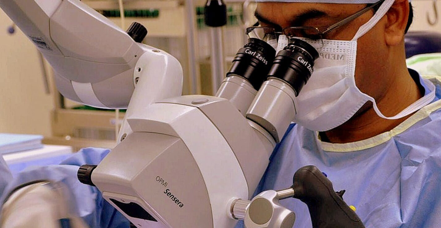
Surgery is performed under general anesthesia in most cases. The primary purpose of surgery is to remove the cholesteatoma so that the ear will dry and the infection will be eliminated. Hearing preservation or restoration is the second goal of surgery.
In cases of severe ear destruction, reconstruction may not be possible. Facial nerve repair or procedures to control dizziness are rarely required. Reconstruction of the middle ear is not always possible in one operation; therefore, a second operation may be performed six to 12 months later. The second operation will attempt to restore hearing and, at the same time, allow the surgeon to inspect the middle ear space and mastoid for residual cholesteatoma.
Surgery can often be done on an out-patient basis. For some patients, an overnight stay is necessary. In rare cases of serious infection, prolonged hospitalization for antibiotic treatment may be necessary. Time off from work is typically one to two weeks.
After surgery, follow-up office visits are necessary to evaluate results and to check for recurrence. In cases where an open mastoidectomy cavity has been created, office visits every few months are needed to clean out the mastoid cavity and prevent new infections. Some patients will need lifelong periodic ear examinations. Cholesteatoma is a serious but treatable ear condition which can be diagnosed only by medical examination. Persistant earache, ear drainage, ear pressure, hearing loss, dizziness, or facial muscle weakness need to be evaluated by an otolaryngologist
Initially, the ear may drain fluid with a foul odor. As the cholesteatoma pouch or sac enlarges, it can cause a feeling of fullness or pressure in the ear, along with hearing loss. An ache behind or in the ear, especially at night, may cause significant discomfort.
Dizziness, or muscle weakness on one side of the face (the side of the infected ear) can also occur. Any or all of these symptoms are good reasons to seek medical evaluation. An ear cholesteatoma can be dangerous and should never be ignored. Bone erosion can cause the infection to spread into the surrounding areas, including the inner ear and brain. If untreated, deafness, brain abscess, meningitis, and, rarely, death can occur. (Source: This page is adapted from a brochure published by the American Academy of Otolaryngology – Head and Neck Surgery, Inc.)
A hole or rupture in the eardrum, a thin membrane that separates the ear canal and the middle ear, is called a perforated eardrum. The medical term for eardrum is tympanic membrane. The middle ear is connected to the nose by the eustachian tube, which equalizes pressure in the middle ear. A perforated eardrum is often accompanied by decreased hearing and occasional discharge. Pain is usually not persistent.
The causes of a perforated eardrum are usually from trauma or infection. A perforated eardrum from trauma can occur:
Middle ear infections may cause pain, hearing loss, and spontaneous rupture (tear) of the eardrum, resulting in a perforation. In this circumstance, there maybe infected or bloody drainage from the ear. In medical terms, this is called otitis media with perforation. Symptoms of acute otitis media include a sense of fullness in the ear, diminished hearing, pain, and fever. On rare occasions a small hole may remain in the eardrum after a previously placed pressure-equalizing (PE) tube falls out or is removed by the physician. Most eardrum perforations heal on their own within weeks of rupture, although some may take several months to heal. During the healing process the ear must be protected from water and trauma. Eardrum perforations that do not heal on their own may require surgery.
sually the size of the perforation determines the level of hearing loss – a larger hole will cause greater hearing loss than a smaller hole. The location of the perforation also affects the degree of hearing loss. If severe trauma (e.g., skull fracture) dislocates the bones in the middle ear which transmit sound, or injures the inner ear structures, hearing loss may be severe.
If the perforated eardrum is caused by a sudden traumatic or explosive event, the loss of hearing can be great and tinnitus (ringing in the ear) may be severe. In this case, hearing usually returns partially, and the ringing diminishes in a few days. Chronic infection as a result of the perforation can cause persistent or progressive hearing loss.
Before attempting any correction of the perforation, a hearing test should be performed. The benefits of closing a perforation include prevention of water entering the ear while showering, bathing, or swimming (which could cause ear infection), improved hearing, and diminished tinnitus. It also may prevent the development of cholesteatoma (skin cyst in the middle ear), which can cause chronic infection and destruction of ear structures. If the perforation is very small, an otolaryngologist may choose to observe the perforation over time to see if it will close spontaneously. He or she might try to patch a patient’s eardrum in the office. Working with a microscope, your doctor may touch the edges of the eardrum with a chemical to stimulate growth and then place a thin paper patch on the eardrum. Usually with closure of the tympanic membrane, hearing is improved. Several applications of a patch (up to three or four) may be required before the perforation closes completely. If your physician feels that a paper patch will not provide prompt or adequate closure of the hole in the eardrum, or if paper patching does not help, surgery may be required.
There are a variety of surgical techniques, but most involve grafting skin tissue or Temporalis fascia across the perforation to allow healing. The name of this procedure is called tympanoplasty. Surgery is typically quite successful in repairing the perforation, restoring or improving hearing, and is often done on an outpatient basis.
our doctor will advise you regarding the proper management of a perforated eardrum. . (Source: This page is adapted from a brochure published by the American Academy of Otolaryngology – Head and Neck Surgery, Inc.)
Copyright @ 2023 | All Rights Reserved by : DAS ENT Care