Our knowledge about sinunasal diseases and their management were limited when 25 years ago Professor Messerklinger identified the mucociliary drainage system of the sinuses which changed the entire way of looking at sinus diseases and managing them. Functional Endoscopic Sinus Surgery is a term coined by an American ENT Surgeon, Dr David Kennedy in 1985to describe the diagnosis and treatment of diseases of the nose and sinusesusing endoscopes and CT scans. Kennedy was introduced to sinus endoscopy in Graz, Austria by Professors Messerklinger and Stammberger. FESS is not one operation, but rather a range of diagnostic and treatment procedures carried out with the help of rigid nasal endoscopes.
The rigid nasal endoscope is a small instrument about 4mm in diameter with a light on the end. It is available in a range of angulations to see around corners and with a powerful fibreoptic light source, the surgeon gets detailed close-up views of the internal nose and sinuses. This helps in the diagnosis and planning the surgery. Only a minority of patients with sinus problems need an operation.
Diagnosis of sinus disease without a CT scan is incomplete. Although we can see the narrow areas where the sinuses open into the nose, we can’t usually see inside the sinuses themselves, unless the openings have already been enlarged.
Plain X-rays of the sinuses are a waste of time and money. They do not show the details and are not used much anymore in the evaluation of the sinus diseases. CT scans allow the ENT Surgeon to examine the sinuses in detailed cross-sections. Virtually all the sinuses can be seen and studied individually.
Unlike getting an MRI, it is rare for patients to feel claustrophobic because you are not enclosed within the scanner. The CT scanner is an open machine. The Procedure is painless and takes less than 10 minutes. Injection of contrast medium at the time of scanning is not necessary for routine CT scans.
Nasal endoscopy only helps the ENT surgeon to visualize the inside of the nose and the sinus openings into the nasal cavity. The inside of the sinuses are not seen during endoscopy unless previous surgery has resulted in enlarged openings. The CT scan enables the ENT surgeon to see inside the sinuses at different cross sections viz. Front to back, side to side and top to bottom. A 3D reconstruction is also possible and adds to better evaluation of sinus diseases. The scan provides a road map for the Sinus surgeon and it will not be wise for the surgeon to perform sinus surgery without performing a CT scan if a good outcome of the surgery without complications is expected.Although CT scanning is a great advance, there are important limitations on what the scan can tell us. A scan on its own does not diagnose sinus problems. It doesprovide some additional information. That information must be weighed and interpreted by the ENT specialist, in the light of the history, examination and endoscopic findings.
A scan is a snapshot of the state of the nose and sinuses on the day it is taken. If you recently had a head cold, the soft tissue lining of the nose and sinuses will be swollen and the scan will look abnormal. On the contrary, if you get recurrent episodes of sinusitis, but haven’t had one for a few weeks, your scan might look completely normal. The only thing that will not change is the bone structure, and the relationship of the eye and brain to the sinuses.
The bone structure is important to us if we are considering an operation on the sinuses. People vary. Sometimes the optic nerve takes a short cut through the sphenoid sinus on its way to the brain. It is better to know this in advance. There is a risk of blindness from damaging the stray optic nerve during surgery.
MRI scans are less useful than CT for most sinus problems, because they don’t show fine bone detail. Some abnormalities in the sinuses may be detected as incidental findings on MRI scans carried out for other reasons, even when there is no real problem. Unless you are getting symptoms of sinus problems, we don’t normally need to do anything about a sinus abnormality on an MRI scan. An MRI is helpful in some rare cases of sinus tumours.
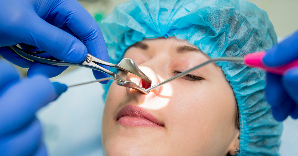
FESS operations are usually done under general anaesthesia (fully asleep) in the operating theatre. The anaesthetist usually puts you to sleep by injection. You will be asleep within a few seconds. The anaesthetist then puts a plastic tube through your mouth into the trachea (windpipe) so that you can breathe during the operation.
In FESS there are no external cuts, the surgery is done through the nostrils. The endoscope attached to a camera is passed through the nose and the interior of the nose can be visualized on the monitor. Specially designed fine bone cutting instruments are then passed through the nostril to enlarged the sinus openings and remove swollen mucosa or polyps blocking them. Other procedures such as septoplasty and LASER vapourization of inferior turbinates are often done at the same time as FESS. Sometimes stitches are applied in septoplasty operation which will be internal and self-dissolving. Most FESS operations take less than an hour to perform. At the end of the operation a sponge dressing will be inserted into each nostril which will be removed after 24 hours.
The success rate of surgery depends on the type of disease treated. Generally people with nasal allergy and polyps will not be cured of their underlying allergy but will get rid of many of their symptoms with certain medications continued in the post operative period. People whose problems are related to mechanical obstruction to mucous flow will be completely cured of their symptoms after surgery. In general between 80 and 90% of patients get great relief of their symptoms and are very pleased with the results of FESS.
Where the principal symptoms are blockage of the nose, facial pain or headache, the results are good. If the main complaint is of post-nasal drip, the results are less encouraging and only around 60% of patients experience meaningful improvement. In patients who present with reduced sense of smell will benefit from FESS, but this may take several months and further post-operative treatment with nasal steroids.
Nasal and sinus operations are very safe procedures today. But no operation is totally risk free. A general anaesthetic carries a minimal risk with consultant anaesthetists using modern drugs and monitoring equipment. There is a risk of excessive bleeding, either during or up to two weeks after the operation. About 2% of patients may need a second operation to control bleeding, readmission to hospital, or a blood transfusion. If you are having a septoplasty (straightening of the central partition between the nostrils) there is a small risk of cosmetic deformity. Operations done for Ethmoidal polyposis carry a small risk of damage to the surrounding structures, including the eyes and the brain which may result in blindness, or a leakage of CSF and Meningitis (extremely rare with experienced surgeons).
Copyright @ 2023 | All Rights Reserved by : DAS ENT Care